Figure 1. [The normal human retina fundus]. - Webvision - NCBI
Por um escritor misterioso
Descrição
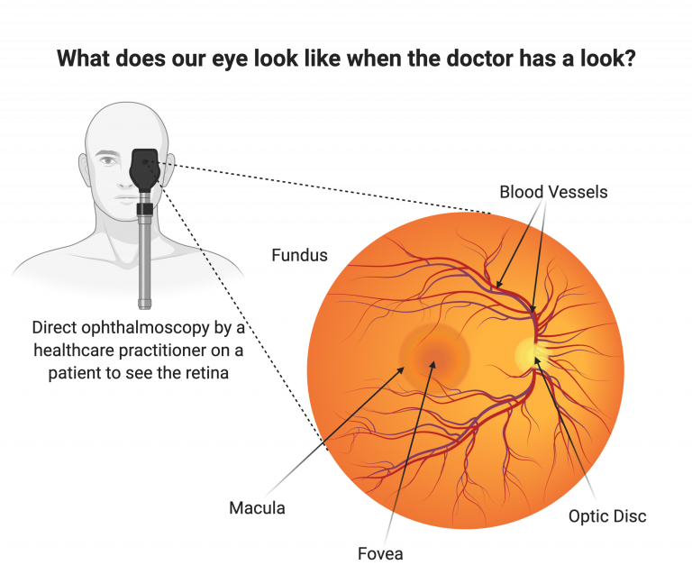
The normal human retina fundus photo shows the optic nerve (right), blood vessels and the position of the fovea (center).
![Figure 1. [The normal human retina fundus]. - Webvision - NCBI](http://webvision.med.utah.edu/imageswv/FlorretBV.jpg)
Simple Anatomy of the Retina by Helga Kolb – Webvision
![Figure 1. [The normal human retina fundus]. - Webvision - NCBI](https://www.ncbi.nlm.nih.gov/books/NBK11556/bin/factsf5.gif)
Facts and Figures Concerning the Human Retina - Webvision - NCBI Bookshelf
![Figure 1. [The normal human retina fundus]. - Webvision - NCBI](https://eophtha.com/images/uploads/15974739483541657755f37849ce2a63.jpg)
Anatomy of Retina
![Figure 1. [The normal human retina fundus]. - Webvision - NCBI](http://eyerounds.org/atlas/LARGE/Normal-fundus-LRG.jpg)
Atlas Entry - Situs Inversus of the Retinal Vessels
![Figure 1. [The normal human retina fundus]. - Webvision - NCBI](https://www.researchgate.net/publication/333702798/figure/fig1/AS:771957922480128@1561060519865/The-human-retina-with-different-stages-of-NPDR-a-normal-retina-and-its-main-components.png)
The human retina with different stages of NPDR: a normal retina and its
![Figure 1. [The normal human retina fundus]. - Webvision - NCBI](https://onlinelibrary.wiley.com/cms/asset/f3b11e32-612e-4803-83bd-bb83b8c9678b/aos14249-fig-0012-m.jpg)
Subretinal surgery: functional and histological consequences of entry into the subretinal space - Sørensen - 2019 - Acta Ophthalmologica - Wiley Online Library
![Figure 1. [The normal human retina fundus]. - Webvision - NCBI](https://www.cell.com/cms/attachment/75170c2e-8c9f-41dc-bb88-3db83ce1f63c/gr3_lrg.jpg)
Suprachoroidal and Subretinal Injections of AAV Using Transscleral Microneedles for Retinal Gene Delivery in Nonhuman Primates: Molecular Therapy - Methods & Clinical Development
![Figure 1. [The normal human retina fundus]. - Webvision - NCBI](https://www.researchgate.net/publication/7614730/figure/fig3/AS:667850092081169@1536239278615/A-normal-fundus-and-those-of-premature-infants-with-ROP-A-Shows-the-normal-vascular.png)
A normal fundus and those of premature infants with ROP. A. Shows the
![Figure 1. [The normal human retina fundus]. - Webvision - NCBI](https://journals.sagepub.com/cms/10.1177/1535370218816517/asset/images/large/10.1177_1535370218816517-fig5.jpeg)
Functional optical coherence tomography of retinal photoreceptors - Xincheng Yao, Taeyoon Son, Tae-Hoon Kim, Yiming Lu, 2018
![Figure 1. [The normal human retina fundus]. - Webvision - NCBI](https://www.ncbi.nlm.nih.gov/books/NBK11533/bin/sretinaf16.gif)
Simple Anatomy of the Retina - Webvision - NCBI Bookshelf
![Figure 1. [The normal human retina fundus]. - Webvision - NCBI](https://media.springernature.com/m685/springer-static/image/art%3A10.1186%2Fs13024-023-00655-y/MediaObjects/13024_2023_655_Fig4_HTML.png)
Retinal ganglion cell repopulation for vision restoration in optic neuropathy: a roadmap from the RReSTORe Consortium, Molecular Neurodegeneration
![Figure 1. [The normal human retina fundus]. - Webvision - NCBI](https://www.ncbi.nlm.nih.gov/books/NBK11533/bin/muller.gif)
Simple Anatomy of the Retina - Webvision - NCBI Bookshelf
![Figure 1. [The normal human retina fundus]. - Webvision - NCBI](https://www.ncbi.nlm.nih.gov/books/NBK1222/bin/retinoschisis-Image001.jpg)
Figure 1. [Fundus photo of a male]. - GeneReviews® - NCBI Bookshelf
de
por adulto (o preço varia de acordo com o tamanho do grupo)